-
Call Now
1800-102-2727


Prokaryotic Cell Definition and Examples
Introduction:
- Prokaryotic cells are generally smaller and simpler than the eukaryotic cells.
- The most conspicuous feature that distinguishes them from eukaryotic cells, is their lack of nucleus and membrane bound organelles.
- The difference in the nuclear structure is the basis of terms prokaryotic ( primitive nucleus) and eukaryotic (well developed nucleus).
- They inhabit a range of habitats and are highly evolved metabolically, since they can meet their energy requirement using very few and simple molecules.
Topics covered:
- Structure of prokaryotic cell/ Bacterial cell
- Cell envelope (Glycocalyx, cell wall and cell membrane) and its modifications
- Gram+ and Gram- bacteria
- Membranous extensions- mesosomes and chromatophores
- Surface extensions and their functions - flagella, fimbriae and pili
- Ribosomes & inclusion bodies
- Plasmids
Structure of prokaryotic cell/ Bacterial cell
Introduction:
- A prokaryotic cell consists of a cell envelope, cytoplasm with ribosomes, nucleoid, plasmids, inclusion bodies, flagella, pili and fimbriae.
- In a typical prokaryotic cell, there is no well defined nucleus and the genetic material is contained in a nucleoid which is not enclosed within the nuclear membrane.
- The DNA is a single circular chromosome and apart from it there is a smaller circular DNA called plasmid.
- Plasmid is capable of self replication and this property is useful in bacteria for developing resistance to antibiotics.
- The prokaryotic cell further contains ribosomes and some special structures called cell inclusions.
Detailed explanation:
- Prokaryotic cell: Cell that bears naked genetic material i.e. nucleus without envelope is known as prokaryotic cell. This group is represented by Bacteria, Blue Green Algae (Cyanobacteria), Mycoplasma.|

- They have the following characters:
1. Like eukaryotic cells, they are different in shape and size but smaller than eukaryotes and divide rapidly.
2. Naked genetic material is called genophore / Nucleoid.
3. Genomic DNA is circular and naked (without histone protein) and termed as a single chromosome.
4. Beside genomic DNA, small circular DNA is also present in many bacteria called plasmid which make them antibiotic resistant, regulates some phenotypes and also responsible for bacterial transformation.
5. Most prokaryotic cells, mainly the bacterial cell, have an envelope consisting of three layers, which are tightly bound outer glycocalyx, middle cell wall and innermost cell membrane.
6. Although each layer of the envelope performs a distinct function, they act together as a single protective unit.
7. If these envelopes are stained by Gram stain then they are called Gram positive bacteria while others that don't are called Gram negative bacteria.
8. Glycocalyx, a polysaccharide envelope forms either loose sheath slime layer or thick and tough structure capsule.
9. Cell wall usually consists of peptidoglycan, absent in mycoplasma.
10. The cell wall determines the shape of the cell and provides a strong structural support to prevent the bacteria from bursting or collapsing.
11. Cell membrane consists of lipoprotein. It is a common structure between prokaryotic cells and Eukaryotic cells.
12. The cell membrane is selectively permeable in nature and interacts with the outside world.
13. Essential infoldings of Plasma membranes towards cytoplasm are called Mesosomes. They can be in the form of the Vesicles Tubules and Lamellae.
14. These help in:
a. Cell wall formation
b. DNA replication and distribution to daughter cells
c. Respiration (analogous to mitochondria)
d. Secretion of processes (to increase the surface area of the plasma membrane and enzymatic content).
15. In cyanobacteria, there are other membranous extensions into the cytoplasm called chromatophores that contain pigments (analogous to chloroplast).
16. Cell walls form some filamentous extensions called flagellum. It consists of filament, hook and basal body and helps in locomotion.
17. Besides flagella, Pili and Fimbriae are also surface structures of the bacteria but do not play a role in motility. The pili are elongated tubular structures made of a special protein pilin. The fimbriae are small bristle-like fibers sprouting out of the cell that provide attachment to substratum or host tissue.
18. In prokaryotes, ribosomes are associated with the plasma membrane of the cell. They are about 15 nm by 20 nm in size. 70S type of ribosomes are found in prokaryotic cells. Its two subunits are 50S and 30S. Several ribosomes are joined with mRNA to form polysome or polyribosome for efficient conduction of protein synthesis.
19. Reserved food materials are stored in cytoplasm in the form of non-living inclusion bodies. Inclusion bodies are not bound by any membrane system and lie free in the cytoplasm.
20. Photosynthetic bacteria like green and purple sulphur bacteria show the presence of gas vacuoles which helps to provide buoyancy.
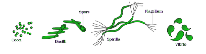
21. The bacteria may exhibit in various shapes- coccus (spherical), bacillus (rod shaped), spirillum (spiral shaped) and vibrio (comma shaped).
Cell envelope ( Glycocalyx, cell wall, and cell membrane ) and its modifications.
Introduction:
- Most prokaryotic cells, particularly the bacterial cells, possess a cell envelope that is chemically complex.
- The cell envelope consists of a tightly bound three layered structure i.e., the outermost glycocalyx followed by the cell wall and then the plasma membrane.
- Although each layer of the cell envelope performs a distinct function, they act together as a single protective unit.
- Bacteria can be classified into two groups on the basis of the differences in the cell envelopes and the manner in which they respond to the staining procedure developed by Gram i.e. Gram positive bacteria (those that take up the gram stain) and Gram negative bacteria ( those which cannot retain the gram stain).
Detailed explanation:
1. Glycocalyx:
- It lies outside the cell wall.
- Glycocalyx differs in composition and thickness among different bacteria. It could be a loose sheath called the slime layer in some, while in others it may be thick and tough, called the capsule.
- It is a thick, high-molecular-weight secretory substance that is present on the external surface of the cell wall of the bacterial cell.
- This structure is composed of either polysaccharides or polypeptides or both.
- The glycocalyx may be thick or thin, rigid or flexible, depending on their chemical nature.
- The terms capsule and slime layer is frequently used to describe glycocalyx layers. The rigid layers are organized in a tight matrix called a capsule.
- The structure of the glycocalyx, which can be deformed easily and can show its attachment with the bacterial cell wall, is known as the slime layer.
- Capsules of the bacterial cell are composed of polysaccharides.
- However, the capsules of some bacilli (Bacillus anthracis) consist of polypeptides, mainly poly-D-glutamic acid.
- The presence of a capsule in the bacterial cell makes it resistant to phagocytosis.
- Hence, it enhances the virulence or the pathogenicity of the bacterial cell.
- It gives a sticky character to the cell.
- It is not essential for the survival of bacteria but performs various secondary functions such as-
i. Protection from desiccation
ii. Protection from toxic chemicals and drugs
iii. Virulence
iv.Protection from phagocytes
2. Cell Wall:
- Bacterial cells almost always are bounded by a chemically complex cell wall.
- The cell wall protects bacteria against osmotic lysis.
- The cell wall is chemically composed of peptidoglycans (also termed as murein). Peptidoglycans are unique to bacterial cells.
- The peptidoglycan present in the cell wall of the Gram-positive bacteria is composed of a single thick homogeneous layer ( 20 to 80 nm), outside the plasma membrane.
- In contrast, the Gram-negative bacterial cell wall consists of a 2 to 7 nm thick peptidoglycan layer covered by a 7 to 8 nm thick outer membrane.
- Peptidoglycan is a polymer containing two sugar derivatives N-acetylmuramic acid (NAM) and N-acetylglucosamine (NAG) joined through an β -1,4 glycosidic bond.
- NAM is NAG with lactic acid attached by an ether linkage.
- A peptide chain of four alternating L- and D- amino acids called tetrapeptide is connected to the carboxyl group of the NAM.

3. Plasma membrane:
- It is the innermost component of the cell envelope.
- It is a selectively permeable covering of the cytoplasm.
- The bacterial membrane has a structure similar to that of a typical membrane.
- It consists of phospholipid bilayer and proteins of several types( extrinsic, intrinsic, transmembrane).
- The bacterial membrane is metabolically active and participates in respiration, synthesis of cell wall components, etc.
Gram-positive and Gram-negative bacteria
Introduction:
- The bacterial cell is composed of a thick cell wall that responds to the gram staining technique.
- On the basis of the response towards gram stain, the bacterial cell is categorized as gram-positive and gram-negative bacteria.
- This staining involved the use of crystal violet or methylene blue as the primary color.
- This gram stain was discovered by the Christian Gram in 1884.
Definition and characteristics features:
- Gram staining is a technique that has a wider application in estimating the kind of bacteria such as gram-positive or gram-negative bacteria at the site of infection.
- The cells which retain the blue or purple color when treated with the gram stain are considered as the Gram (+) bacteria. For example, Bacillus subtilis.
- The cells which do not retain any gram stain and become colorless are termed as Gram (-) bacteria. For example, Escherichia coli.
Detailed explanation:
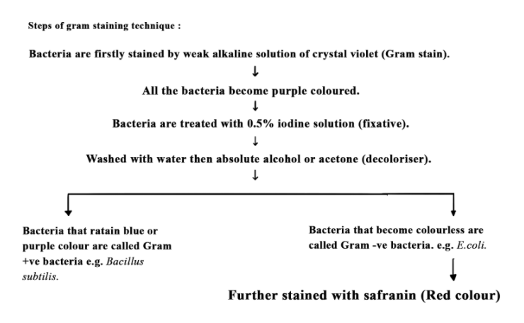
- The staining of the bacterial cell occurs with the help of a weakly alkaline solution of crystal violet or gentian violet.
- This cell adds blue-violet color to its cell coat.
- Further, they are treated with 0.5% iodine solution followed by washing with water and then absolute alcohol or acetone.
- The cells which retain the blue or purple color when treated with the gram stain are considered as the Gram (+) bacteria. For example, Bacillus subtilis.
- The cells which do not retain any gram stain and become colorless are termed as Gram (-) bacteria. For example, Escherichia coli.
- The gram-negative bacteria are generally counterstained with the help of saffarine.
- The gram stain is washed away from the gram-negative bacteria due to the presence of high lipid content in the cell wall which gets dissolved in organic solvents like acetone.
Differences between Gram-positive and Gram-negative Bacteria
| Gram-positive bacteria | Gram-negative bacteria |
|---|---|
| The bacterial cells retain blue or purple color after gram staining when washed with acetone. | The bacteria do not retain the stain when washed with absolute alcohol. |
| This cell shows the absence of an outer membrane and is considered as a single-layered cell. | The cells show the presence of the outer membrane and are composed of two layers. |
| The thickness of the wall is more. | The thickness of the wall is less as compared to gram-positive bacteria. |
| The bacterial wall contains less amount of lipid percentage(2-4%). | The bacterial wall contains more lipid percentage (20-30%). |
| Mesosomes are more prominently present. | Mesosomes are generally absent. |
| The flagellum has two rings of swelling in the structure of the basal body. | The flagellum has four rings of swelling in the structure of the basal body. |
| The wall is composed of Murein or mucopeptide content approx. 70-80%. | The wall is composed of Murein or mucopeptide content approx. 10-20%. |
| The hydrophilic channels such as porins are absent. | The hydrophilic channels such as porins are present on the outer membrane of the cell wall. |
| Very few pathogenic bacteria belong to the Gram-positive group. | More of the pathogenic bacteria belong to the Gram-negative group. |
| TThese bacteria are more susceptible to antibiotics. | These bacteria are more resistant to antibiotics. |
| The cell wall contains teichoic acids. | The cell wall does not contain teichoic acids. |
Note- Teichoic acid in Gram positive bacteria :
- Many Gram-positive bacteria have acidic substances called teichoic acids in their cell wall.
- Teichoic acids are polyphosphate polymers (a polymer of glycerol or ribitol joined by phosphate) bearing a strong negative charge.
- This acid is linked covalently with the N-acetylmuramic acid of the peptidoglycan layer and can also show attachment with the lipids present in the plasma membrane.
- The combination of teichoic acids and lipids is referred to as lipoteichoic acids. Teichoic acids are absent in Gram-negative bacteria.
- The main function of teichoic acids is to provide rigidity to the cell wall by attracting cations such as magnesium and sodium.
Membranous extensions:
Introduction:
- The membrane of the bacterial cell undergoes internal invasion which is known as membranous extensions.
- There are two types of membranous extensions that are generally studied in bacterial cells.
- These are mesosomes and chromatophores.
- Mesosome is the characteristic circular to the villiform specialization of cell membrane of bacteria that develops as an ingrowth of plasma membrane.This structure develops as an ingrowth of the plasma membrane and can be vesicular, tubular, or lamellate.
- Chromatophores are the membrane extensions in photosynthetic forms such as purple and green sulphur bacteria which possess photosynthetic pigments.
Detailed explanation:
A. Mesosomes
- A special membranous structure is the mesosome which is formed by the extensions of plasma membrane into the cell.
- These extensions are in the form of vesicles, tubules and lamellae.
- It is of two types:
(i) Septal mesosome
(ii) Lateral mesosome
(i) Septal mesosome:
- The septal mesosome is responsible for connecting the nucleoid region of the cell with that of the plasma membrane.
- The replication of the nucleoid region in the bacterial cell involves the functioning of the septal mesosome by providing points of attachment to the replicated ones.
- It is involved in the formation of the septum.
- At the time of cell division, the plasma region grows in the region where the septal mesosome is present so that it provides membranes for rapid elongation.
(ii) Lateral mesosome:
- Unlike the septal mesosome, the lateral mesosome is not connected with the nucleoid region.
- This structure consists of respiratory enzymes and is also considered to be the equivalent of the mitochondria of eukaryotes.
- It increases the surface area of the plasma membrane and enzymatic content.
Functions of mesosome:
- This structure is responsible for the formation of the cell walls.
- It is also involved in the function of DNA replication along with its distribution to daughter cells.
- This is also involved in the process of respiration, secretion processes, to increase the surface area of the plasma membrane and enzymatic content.
Chromatophores:
- In some forms of photosynthetic bacteria, a chromatophore is a coloured, membrane-associated vesicle used to perform photosynthesis.
- The photosynthetic pigments present in the chromatophores are bacteriochlorophyll and carotenoids.
- In Rhodospirillum rubrum, a purple bacteria, the light-harvesting proteins are internal to the chromatophore membranes.
- Whereas in green sulphur bacteria, the light harvesting proteins are arranged in specialized antenna complexes called chlorosomes.
Surface extensions and their functions - flagella, fimbriae, and pili
Introduction:
- Bacterial cells may consist of certain surface extensions which include- Flagella, pili and fimbriae.
- Bacterial flagella are long, filamentous surface appendages and are the extension of the cell wall of the bacteria. They help in motility.
- The term pilli (singular: pilus) is used to describe the thin, hairlike appendages on the surface of bacteria that aids in sexual recombination(conjugation).
- Fimbriae may be present in both Gram-negative and Gram-positive bacteria. They are small bristle-like fibers sprouting out of the cell that provide attachment to substratum or host tissue.
Detailed explanation:

A. Flagella:
- The gram-positive and gram-negative bacteria may show the presence of the motile appendage of flagella.
- Bacterial flagella are long, thin, filamentous extensions of the cell wall.
The gram-negative bacterium such as E. coli contains flagella which consists of three parts:
- The long filament occurs in the exterior region of the cell surface.
- The hook structure is present at the end region of the filament.
- The basal body is the structure that is associated with the hook and together supports the motion of the flagellum.
- Some bacteria have sheaths surrounding their flagella.
- The flagellar filament is made up of flagellin protein.
- In filament, the flagellin proteins are the globular proteins which are assembled to form a cylindrical structure with a hollow core. The flagellin molecules constituting the flagellar filament are arranged in the spiral rows.
- Hook is the curved tubular structure which connects the filament with the basal body and is the thickest part of the flagellum.
- The basal body is rod like structure inserted in the cell envelope.
- The basal bodies of typical Gram-negative bacteria have two pairs of rings that are L and P rings in the cell wall and S and M rings embedded in the cell membrane.
- Gram positive bacteria consist of only a single pair of rings (S and M rings) embedded in the cell membrane.
- A proton pump or stator and a molecular motor or rotator occur at the bottom of the basal body.
- The bacterial flagella perform rotation type movement that brings about backward pushing of the water. It results in the bacterium moving forward.
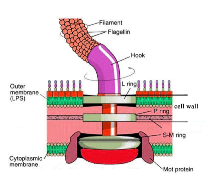
- Note- Earlier, it was thought that the basal body comprises four rings M (for Membrane), S (for Supramembranous), P (for Peptidoglycan), and L (for Lipopolysaccharide).
- Later, it was found that both the M- and S-rings (now called the MS-ring) comprise different domains of the same protein.
- Therefore, they function as a unit.
B. Pili:
- The term pill (singular: pilus) is used to describe the thin, tubular, hairlike appendages on the surface of bacteria.
- Pili are commonly found in Gram-negative bacteria.
- Proteins that form pili are referred to as pilin.
- It plays an important role in the process of conjugation( sexual recombination).
- It allows the transfer of genetic material from a donor bacterial cell having fertility factor to another cell that acts as recipient and lacks the fertility factor by the formation of conjugation pili. Hence, it is also called the sex pili.
C. Fimbriae:
- Fimbriae are small, bristle-like sprouting from the cell surface in large numbers.
- They are present in both Gram-negative and Gram-positive bacteria.
- Fimbriae are not involved in motility but help the bacteria to stick to one another or help them attach to the substratum.
Differences between pili and fimbriae :
| Pili | Fimbriae |
|---|---|
| Pili Present in Gram-negative bacteria. | Fimbriae are present in both Gram-negative and Gram-positive bacteria. |
| Required for bacterial conjugation. | Used for attachment of cells to the surface. |
| It is present quite less in number per cell. | Number up to several hundred per cell. |
| Long, tubular structure. | Short, bristle like solid structure. |
Ribosomes & Inclusion bodies
Introduction:
- The bacterial cytoplasm contains 70S ribosomes and inclusion bodies such as cyanophycin granules, glycogen, carboxysomes, polyphosphate granules.
Ribosomes:
- The prokaryotic cell consists of 70S types of ribosomes which are composed of larger 50S and smaller 30S subunits.
- These are the small membrane-less structures present in the cytoplasm that are the ribonucleoprotein entities.
- These are either associated with the plasma membrane or are freely present in the cytoplasm.
Inclusion bodies:
- Reserve material in prokaryotic cells are stored in the cytoplasm in the form of inclusion bodies. These are not bound by any membrane system and lie free in the cytoplasm.
Detailed explanation:
A. Ribosomes:
- Ribosomes are the small membrane-less structures present in the cytoplasm that are the ribonucleoprotein entities.
- In prokaryotes, ribosomes are associated with the plasma membrane of the cell or occur free in the cytoplasmic matrix.
- Bacterial 70S ribosomes differ from eukaryotic 80S ribosomes present in cytosol in the number of proteins and rRNA molecules they contain.
- The subunits of a 70S ribosome are a small 30S subunit containing (16S rRNA) and a larger 50S subunit-containing (23S and 5S rRNA).
- The bacterial ribosome is composed of two subunits- larger 50S and smaller 30S.
- Ribosomes are the site of protein synthesis.
- Several ribosomes may attach to a single mRNA and form a chain called polyribosomes or polysomes.
- The ribosomes of a polysome translate the mRNA into proteins.
B. Inclusion bodies:
- Inclusion bodies are nonliving substances present in the cytoplasm and usually consist of reserved food material, inorganic inclusions and gas vacuoles.
- They are also known as ergastic substances.
- Cell inclusion bodies present in bacteria are cyanophycin granules, glycogen, carboxysomes, inorganic inclusions (polyphosphate granules, sulphur granules, etc.), and gas vacuoles.
i. Reserved food :
- Cyanophycean starch or the -granules along with -granules or the lipid β-globules and cyanophycin or the protein granules are the types of food reserve found in the blue green algae.
- In bacteria, the starch is replaced by glycogen which is a polymer of glucose and is present in the form of granules in the cytosol.
- Carboxysomes are polyhedral inclusion bodies that contain the CO2 fixation enzymes ribulose-1,5-bisphosphate carboxylase (RuBisCO).
- These structures are found in photosynthetic forms.
ii. Inorganic inclusions :
- They include- volutin granules, sulphur granules, iron granules, etc.
- Polyphosphate granules (or volutin granules) are reserve forms of inorganic phosphate (polyphosphate) present in the cytoplasm of some bacteria.
- Sulphur granules are found in bacteria found in sulphur rich medium which picks up hydrogen sulphide for obtaining the reducing power in photosynthesis.
- The bacteria which metabolizes iron compounds for obtaining energy show the presence of iron granules.
iii. Gas vacuole:
- They are gas storing vacuoles found in cyanobacteria, purple and green bacteria, and a few other planktonic forms.
- A gas vacuole does not have any covering of its own.
- It consists of a variable number of cylindrical gas vesicles which is surrounded by a single non-unit, protein-membrane.
- The membrane is impermeable to water but is permeable to atmospheric gases.
- Gas vacuoles protect the bacteria from harmful radiations and provide buoyancy.
Plasmids:
Introduction:
- Plasmids are the extra-chromosomal segments of the ds DNA ( double-stranded deoxyribonucleic acid).
- These are the self-replicating, circular, and naked DNA structures.
- The plasmid is independent and not part of the nucleoid region of the bacterial cell.
- Some of them contain important genes like fertility factors, resistance factors, and colicinogenic factors.
- These plasmids are widely used in the methods of genetic engineering.
Definition and characteristics features:
- Plasmids are the autonomous self-replication strands of DNA that act as an extrachromosomal genetic element in bacteria.
- The use of plasmid occurs in genetic engineering technology.
- Plasmid shows replication similar to the bacterial chromosomes. They contain the ORI site (Origin of replication) which allows the bi-directional replication of the plasmid.
- The average number of plasmid molecules given in the bacterial cell is known as the copy number.
- This number of copies is randomly observed after the cell division.
- Plasmids carry genes for managing their own lifecycles and some plasmids carry genes that affect the properties of the host cell.
Detailed explanation:
- There are different types of plasmids observed depending upon the functionality-
Different types of plasmid:
A. F-plasmid (Fertility factor plasmid):
- The F-plasmid is an example of the self-transmissible plasmid.
- Bacteria that possess a copy of F plasmid are called F+strain of bacteria whereas the bacterial strain which does not possess the copy of F- plasmid are called F- strain of bacteria.
- Here, the F-plasmid stands for Fertility factor plasmid which is a conjugative plasmid containing genetic information that codes for pilin, used to make sex pilus necessary for conjugation.
- During conjugation, a mode of sexual recombination, F-factor or the fertility factor is transferred from the donor F+ strain to the recipient F- strain by conjugation tube or sex pili.
- One bacterium serves as the donor of the genetic material (carrying a DNA sequence called the fertility factor, or F-factor) and the other bacterium serves as the recipient (showing the absence of F factor), during conjugation.
- A plasmid that can exist autonomously in a cell or can integrate itself into the bacterial chromosome is termed as episome.
- The F- plasmid is an episome.
- A bacterial cell that contains an F-plasmid integrated with the bacterial chromosome is considered as an Hfr (High-frequency recombination) cell.
- Integration involves homologous recombination between two covalently closed circular DNA molecules forming one circular molecule containing both of the original DNA structures.

B. R-plasmid (resistance plasmid):
- R-plasmids make the host cell resistant to one or more antibiotics.
- They typically have genes that code for enzymes capable of destroying or modifying antibiotics.
- Some R- plasmids have only a single antibiotic resistance whereas others have many. Most R-plasmids are mobilizable plasmids.
C. Col (colicinogenic) plasmids:
- Col plasmids synthesize proteins termed as bacteriocin that prevent the growth of susceptible bacterial strains that do not contain a Col plasmid.
- Bacteriocins are bactericidal proteins that inhibit the growth of other strains of the same species as well as other closely related species.
- Bacteriocins are chemically peptides that have the property of killing other bacteria but themselves remain unaffected.
- There are many types of colicins, each designated by a letter (for example, colicin B) and each having a particular mode of inhibition of sensitive cells.
The names of some common plasmids and their functions:
| Plasmid | Gene function |
|---|---|
| Resistance | Resistance against antibiotics. |
| Fertility. | The DNA transfer between the bacterial species (from donor to recipient cells) through the process of conjugation. |
| Killer | The toxin is released to kill other bacteria. |
| Degradative | Enzymes for the metabolism of unusual molecules. |
| Virulence | Pathogenicity |
Frequently asked questions (FAQ)
Q1. Discuss about the cell envelope of the bacterial cell?
Ans:
- The cell envelope is a chemically complex structure surrounding the most prokaryotic cells, particularly the bacterial cells.
- The cell envelope consists of a tightly bound three layered structure that is the outermost glycocalyx followed by the cell wall and then the plasma membrane.
- These layers together form a single protective unit.
Q2. What is the difference between the gram-positive and gram-negative bacteria ?
Ans: The differences between Gram-positive and Gram-negative Bacteria:
Gram positive bacteria:
- The bacterial cells retain blue or purple color after gram staining when washed with acetone.
- This cell shows the absence of an outer membrane and is considered as a single-layered cell.
- The thickness of the wall is more.
- The bacterial wall contains less amount of lipid percentage.
- Mesosomes are more prominently present.
- The flagellum has two rings of swelling in the structure of the basal body.
- The wall is composed of Murein or mucopeptide content approx. 70-80%.
- The hydrophilic channels such as porins are absent.
- Very few pathogenic bacteria belong to the Gram-positive group.
- These bacteria can be destroyed with the action of antibiotics
- The cell wall contains teichoic acids.
Gram negative bacteria:
- The bacteria do not retain the stain when washed with absolute alcohol.
- The cells show the presence of the outer membrane and are composed of two layers.
- The thickness of the wall is less as compared to gram-positive bacteria.
- The bacterial wall contains more lipid percentage (20-30%).
- Mesosomes are generally absent.
- The flagellum has four rings of swelling in the structure of the basal body.
- The wall is composed of Murein or mucopeptide content approx. 10-20%.
- The hydrophilic channels such as porins are present on the outer membrane of the cell wall.
- More of the pathogenic bacteria belong to the Gram-negative group.
- These bacteria cannot be destroyed with the action of antibiotics and hence, it is more resistive in nature.
- The cell wall does not contain teichoic acids.
Q3. Explain the gram staining technique and how the bacteria can be classified on its basis?
Ans:
- This gram stain was discovered by the Christian Gram in 1884.
- This staining involved the use of crystal violet or methylene blue as the primary color.
- On the basis of the response towards gram stain, the bacterial cell is categorized as gram-positive and gram-negative bacteria.
- The cells which retain the blue or purple color when treated with the gram stain are considered as the Gram (+) bacteria. For example, Bacillus subtilis.
- The cells which do not retain any gram stain and become colorless are termed as Gram (-) bacteria. For example, Escherichia coli.
Q4. What are mesosomes and how are they analogous to the mitochondria of eukaryotes??
Ans:
- Mesosome is the characteristic circular to the villiform specialization of cell membrane of bacteria that develops as an ingrowth of plasma membrane.
- These membranous extensions are present in the form of vesicles, tubules, and lamellae.
- This structure is responsible for the formation of the cell walls.
- It is also involved in the function of DNA replication along with its distribution to daughter cells.
- It is also involved in secretion processes.
- This structure consists of respiratory enzymes and is also considered to be the equivalent of the mitochondria of eukaryotes as it increases the surface area of the plasma membrane and enzymatic content.
Q5. Describe the types of mesosomes and its importance?
Ans:
- Mesosome is the special membranous structure that is formed by the extensions of the plasma membrane into the cell.
- It is of two types:
(i) Septal mesosome
(ii) Lateral mesosome
(i) Septal mesosome:
- The septal mesosome is responsible for connecting the nucleoid region of the cell with that of the plasma membrane.
- The replication of the nucleoid region in the bacterial cell involves the functioning of the septal mesosome by providing points of attachment to the replicated ones.
- It is involved in the formation of the septum.
- At the time of cell division, the plasma region grows in the region where the septal mesosome is present so that it provides membranes for rapid elongation.
(ii) Lateral mesosome:
- Unlike the septal mesosome, the lateral mesosome is not connected with the nucleoid region.
- This structure consists of respiratory enzymes and is also considered to be the equivalent of the mitochondria of eukaryotes.
- It increases the surface area of the plasma membrane and enzymatic content.
Q6. Define chromatophore and its functions.?
Ans:
- In some forms of photosynthetic bacteria, a chromatophore is a coloured, membrane-associated vesicle used to perform photosynthesis.
- The photosynthetic pigments present in the chromatophores are bacteriochlorophyll and carotenoids.
- In Rhodospirillum rubrum, a purple bacteria, the light-harvesting proteins are internal to the chromatophore membranes.
- Whereas in green sulphur bacteria, the light harvesting proteins are arranged in specialized antenna complexes called chlorosomes.
Q7. Explain the surface extensions in bacteria that help in motility?
Ans:
- Bacterial cells may consist of certain surface extensions which include- Flagella, pili and fimbriae.
- Bacterial flagella are long, filamentous surface appendages and are the extension of the cell wall of the bacteria. They help in motility.
- Bacterial flagellum is composed of three parts – filament, hook and basal body. The filament is the longest portion and extends from the cell surface to the outside.
Q8. Explain plasmids and its types?
Ans:
- Plasmids are the extra-chromosomal segments of the ds DNA ( double-stranded deoxyribonucleic acid).
- These are the self-replicating, circular, and naked DNA structures.
- There is a different type of plasmids observed depending upon the functionality which are F plasmid, Col plasmid and R plasmid.
F-plasmid (Fertility Factor plasmid):
- Bacteria that possess a copy of F plasmid are called F+ strain of bacteria whereas the bacterial strain which does not possess the copy of F- plasmid are called F- strain of bacteria.
- Here, the F-plasmid stands for Fertility factor plasmid which is a conjugative plasmid containing genetic information that codes for pilin, used to make sex pilus necessary for conjugation.
- During conjugation, a mode of sexual recombination, F-factor or the fertility factor is transferred from the donor F+ strain to the recipient F- strain by conjugation tube or sex pili.
- One bacterium serves as the donor of the genetic material (carrying a DNA sequence called the fertility factor, or F-factor) and the other bacterium serves as the recipient (showing the absence of F factor), during conjugation.
R-plasmids:
- R- plasmid makes the host cell resistant to one or more antibiotics.
- They typically have genes that code for enzymes capable of destroying or modifying antibiotics.
- Some R- plasmids have only a single antibiotic resistance whereas others have many. Most R-plasmids are mobilizable plasmids.
Col (colicinogenic) plasmids:
- Col plasmids synthesize proteins termed as bacteriocin that prevent the growth of susceptible bacterial strains that do not contain a Col plasmid.
Q9. Which cell inclusions are present in bacterial cells?
Ans:
- Inclusion bodies are nonliving substances present in the cytoplasm and usually consist of reserved food material, mineral matter, etc.
- They are also known as ergastic substances.
- Examples of inclusion bodies present in bacterial cells are cyanophycin granules, glycogen, carboxysomes, polyphosphate granules, and other inorganic inclusions, and gas vacuoles.
Q10. What are gas vacuoles and where are they found?
Ans:
- They are gas storing vacuoles found in cyanobacteria, purple and green bacteria, and a few other planktonic forms.
- A gas vacuole does not have any covering.
- It consists of a variable number of cylindrical gas vesicles which is surrounded by a single non-unit, protein-membrane.
- The membrane is impermeable to water but is permeable to atmospheric gases.
- Gas vacuoles protect the bacteria from harmful radiations and provide buoyancy.


Top Biology Concepts
Important Concept Pages
NEET Related Links
- NEET Exam 2022
- NEET 2022 Exam Dates
- NEET Mock Test
- NEET Answer Key Solutions
- NEET Previous Year Question Papers
- NEET Rank Predictor
- NEET 2022 Admit Card
- NEET Result
- NEET Cut Off
- NEET 2022 Syllabus
- NEET Exam Pattern
- NEET 2022 Eligibility Criteria